96 Hour Chick Embryo Serial Section
Contents • • • • • • • • Introduction Historically, an important way of teaching embryology is by direct observation of histological sections at different stages of development. Even today the best way to observe anatomical development and relationships is by direct observation in serial sections through the embryo and fetus. The sections below are from the original teaching set prepared at UNSW that were digitized and put online in 1996 as a teaching resource. The Stage 13 and 22 images are available in at least two formats, an unlabeled set (image 1 - 49) and a labeled set (image 50 - 98) in a rostro-caudal sequence (head to tail). Stage 22 embryo also has a set of selected images showing more detail of some organ and tissue development.
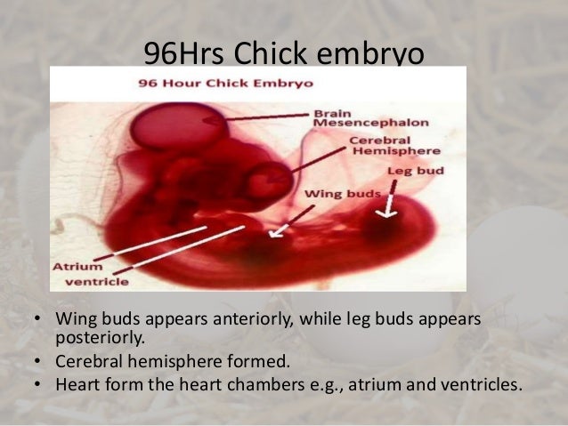
Of early embryo of Penaeus orientalis sections (7 pieces),Development of early Embryo of Chicken w.m. & serial transverse sections(24/48/96 hours).
In addition, there are now 3D animations based upon reconstruction of the embryos from these serial sections. 2011 - Selected slides are currently being rescanned at to show specific developmental details. Myscript stylus linux.
Author Comments Start here by looking through the early (week 4) embryo (stage 13) labeled images. It is not so important to identify every single feature, observe what structures are present and where they are in the middle of the embryonic period (week 4).
Then if you like, look through the unlabeled images. What can you now recognise? Next look through the late (week 8) embryo (stage 22) labeled images as you did before. Then look through the same embryo stage selected labeled images. These show organ and tissue development of specific structures by the end of the embryonic period. What has changed?
Finally put the systems together by watching the embryo animations based upon these sections. How different are the embryonic systems to the adult anatomy? More about these.
Links: Carnegie Stage 13 About Stage 13 Embryo Sections - This image is from a serial section of a 6mm CRL pig embryo with some features of the Stage 14 embryo. This embryo is approximately equal to the day 42 human embryo. Use these serial images to identify internal features and relationships that exist within the embryo at this stage. Then compare these images with the later features of the Carnegie stage 22 human embryo.
At about 33 hours after fertilization, the embryo is about 4 mm long and the first flexion of the originally straight embryo starts in the head region. The cranial flexure will be visible a few hours later. At this stage 12 to 13 somites are formed.
The eye vesicles are rather large. The forebrain vesicle or prosencephalon will divide, the midbrain vesicle or mesencephalon remains undivided while the hindbrain vesicle or rhombencephalon will form a series of smaller neuromeres. The sinus rhomboidalis (diamond-shaped) is still present as the only opening of the neural tube and the primitive streak is only rudimentary. The infundibulum (= derived from the diencephalon) appears as a half circular structure at the ventral side of the caudal part of the forebrain. The notochord or chorda dorsalis ends just behind this ventral vesicle. Later on, 36 hours after fertilization, the heart, which has a bilateral origin in the mesodermal layer, is a S-shaped tube which protrudes to the right of the embryo (in upper view).

Outside, in the area vasculosa (= forseen of blood vessels) the formation of blood islands continues. The primitive streak can only still be discerned below the sinus rhomboidalis.
Early embryonic developmental features that are shown on this page: • Whole mount preparation 33 hours () • Whole mount preparation 36 hours () • Cross sections 36 hrs; formation of eye, heart and intestines ( ) Stage 33 hours Information: At about 33 hours after fertilization, the embryo is about 4 mm long and the first flexion of the originally straight embryo starts in the head region and the cranial flexure will be visible a few hours later. At this stage 12 to 13 somites are formed. The eye vesicles are rather large. The forebrain vesicle or prosencephalon will divide, the midbrain vesicle or mesencephalon remains undivided while the hindbrain vesicle or rhombencephalon will form a series of smaller neuromeres. The sinus rhomboidalis (diamond-shaped???) is still present as the only opening of the neural tube and the primitive streak is only rudimentary.
The infundibulum (= derived from the diencephalon) appears as a half circular structure at the ventral side of caudal part of the forebrain. The notochord or chorda dorsalis ends just behind this venral vesicle. Embryology of chicken 33 hours after fertilization: stained whole mout preparation 1 = Proamnion, 2 = Prosencephalon, 3 = Mesencephalon, 4 = Rhombencephalon, 5 = Somite, 6 = Eye vesicle, 7 = Foregut, 8 = Chorda (translucent), 9 = Heart, 10 = Lateral mesoderm, 11 = Spine, 12 = Sinus rhomboidalis, 13 = Primitive streakp, 14 = Blood islands Dorsalview and longitudinalsection at 33 hrs according to Patten Patten, B.M. The Early Embryology of the Chick. Philadelphia: P.